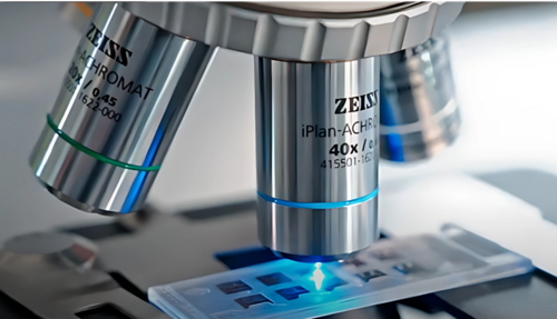
The world of science has long relied on the ability to explore the unseen. From the first rudimentary lenses to today’s advanced imaging systems, microscopy has transformed our understanding of life and its microscopic details. In this blog, we’ll look through the fascinating history of microscopy, highlighting key milestones and focussing on how modern innovations like the EUROPattern and EUROPattern Microscope Live from EUROIMMUN continue to revolutionise the field.
The Early Days: Glass Lenses and Curiosity
The origins of microscopy trace back to the late 16th century, when Dutch spectacle makers Hans and Zacharias Janssen created the first compound microscope. This device used multiple lenses to magnify objects, setting the stage for a new era of scientific discovery. Soon after, Galileo Galilei adapted a similar concept to study celestial bodies, further demonstrating the power of lenses.
In the 1670s, Antonie van Leeuwenhoek, a Dutch scientist and tradesman, took microscopy to new heights. Using his finely crafted single-lens microscopes, he observed and described bacteria, protozoa and sperm cells for the first time. These discoveries marked the beginning of microbiology and laid the groundwork for cellular biology.
The Advent of Modern Microscopy
The 19th century saw significant advancements in optical theory and technology. Carl Zeiss and Ernst Abbe developed precision lenses and illumination systems, drastically improving resolution and contrast. These innovations allowed scientists to observe cellular structures in greater detail, fostering breakthroughs in medicine and biology.
The invention of the electron microscope in the 1930s marked a paradigm shift. Using electron beams instead of light, this technology achieved unprecedented magnification, revealing the intricate details of viruses and molecular structures. From there, microscopy diversified into specialised techniques such as fluorescence microscopy, confocal microscopy and super-resolution microscopy, each designed to uncover specific aspects of the microscopic world.
Microscopy in the Digital Age
The integration of digital technologies has revolutionised microscopy in recent decades. High-resolution imaging, automated sample analysis and data sharing have become integral to modern systems, making microscopy faster and more accessible than ever.
EUROIMMUN’s EUROPattern Technology exemplifies this transformation. Combining automated fluorescence microscopy with sophisticated pattern recognition software, the EUROPattern system provides rapid and precise analysis of immunofluorescence tests. The system eliminates the need for manual evaluation, significantly reducing subjectivity and enhancing diagnostic accuracy.
EUROPattern Microscope Live: A Leap Forward
Building on this innovation, EUROIMMUN introduced EUROPattern Microscope Live (EPML), an advanced system that takes automation to the next level. This cutting-edge microscope integrates live imaging capabilities with real-time processing, enabling on-the-spot evaluation of samples. Its seamless operation allows laboratories to handle high workloads efficiently while maintaining exceptional diagnostic quality.
The EPML system is particularly beneficial for autoimmune and infectious disease diagnostics, where speed and precision are critical. With features such as remote access and intuitive software interfaces, EPML represents a perfect blend of traditional microscopy principles and modern technology, driving progress in clinical diagnostics.
The Future of Microscopy
As we look ahead, the future of microscopy promises even greater innovations. Advances in artificial intelligence, machine learning and nanoscale imaging are set to unveil new dimensions of the microscopic world. Microscopy has come a long way from its humble beginnings, and its evolution continues to shape how we perceive and interact with the world at a cellular level. By integrating tradition with innovation, modern microscopy not only honours its history but also drives us toward a brighter future in science and medicine.
References
- Ball, Clara Sue. "The Evolution of the Microscope." JSTOR Daily. Explores the historical development of microscopy, from early compound microscopes to modern advancements.
- Harvard Science Review. "A History of Microscopy." Details the shift from optical to electron microscopy, highlighting breakthroughs like transmission electron microscopes and scanning tunneling microscopy.
- SpringerLink. "Brief History of Microscopy." Provides an overview of key developments in light, electron, and advanced atomic-level microscopy.


